CBCT Field of View: How to Compare Systems for Your Dental Practice
Field of view, or FOV, describes the volume of anatomy captured in a cone beam CT scan. When you compare CBCT systems, the useful question is not simply “How large is the maximum field?” It is “Which acquisition options and software tools support the cases I expect to evaluate?”
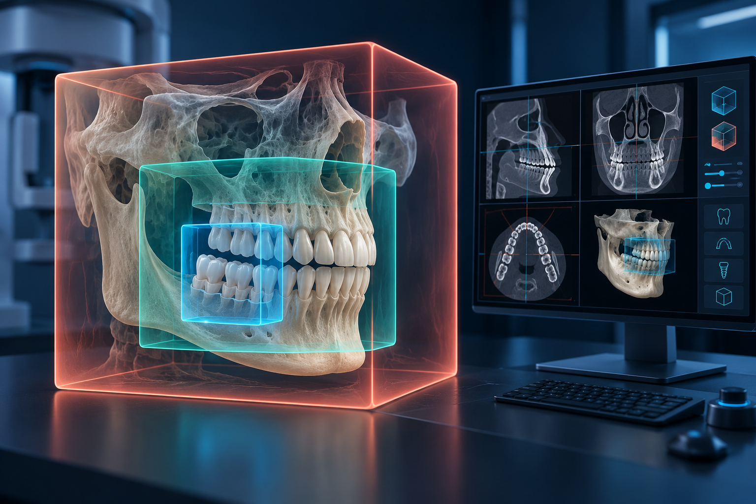
Keep two decisions separate: buying a system with suitable acquisition options, and selecting a justified scan for the individual patient.
Start with anatomy, not small, medium, or large
Manufacturer and distributor labels are not a universal sizing standard. Ask for the actual field dimensions, volume shape, positioning limits, and available smaller acquisitions on the exact model and configuration.
FDA guidance says dental CBCT use should be justified by the clinical question and optimized for the patient, indication, and anatomy being scanned. The FDI’s current radiation-safety policy similarly emphasizes indication-oriented, patient-specific imaging with diagnostic quality. 1
The largest available field is not the setting to use for every patient. Field of view is also not a stand-alone measure of image quality or radiation exposure; protocols, patient factors, acquisition settings, reconstruction, and device design matter.
If your work is localized, test the limited acquisitions
Endodontic questions and localized implant sites may fit limited acquisitions, but procedure count alone does not determine the required anatomy. One implant near a relevant structure may require different coverage from another implant in a different location.
During a demonstration, ask the supplier to show:
- each limited acquisition available on the quoted system;
- the volume dimensions and shape;
- how the patient is positioned;
- how the region is selected and verified;
- how the dataset appears in the viewing software;
- which settings are tied to the protocol.
A machine with a large maximum field may still offer useful smaller acquisitions. Verify that capability on the exact model instead of assuming that software cropping is the same as acquiring a smaller volume.
If you plan full-arch cases, verify the complete coverage
For full-arch implant planning, ask whether the intended arch and relevant surrounding anatomy fit inside the available acquisition. Do not rely on the word “medium.” Request the dimensions and work through a representative demonstration case.
If pterygoid or zygomatic implant planning is within your intended scope, define the required anatomy with the clinician doing that planning. Excluding those procedures does not automatically prove that a particular field is sufficient.
These are system-purchase questions, not fixed prescriptions for how every patient should be scanned.
Do not use airway interest as a shortcut
Broader anatomical assessment may change the coverage a practice wants available. But offering an oral appliance or evaluating an airway does not automatically mean the office needs the largest field or should scan every patient.
The American Academy of Sleep Medicine’s adult diagnostic guideline uses polysomnography or, for appropriate patients, home sleep apnea testing for diagnosis. A CBCT airway image does not replace diagnostic sleep testing. 2
Ask what clinical question the image will answer, which anatomy is required, and whether the exam is justified. Keep equipment capability separate from a patient-level indication.
Test the software alongside the hardware
Khamir recommends evaluating the viewing and planning software before purchase, not only the scanner specifications. 3
Ask to operate the software yourself with a demonstration dataset. Try the tasks your practice expects to perform:
- find and orient the relevant views;
- adjust basic display settings;
- take measurements;
- trace or inspect relevant anatomy where appropriate;
- work through an implant plan if that is part of your workflow;
- export or share the study in the format your referral and lab partners need;
- confirm who can view, plan, report, and administer the system.
Have the supplier identify which functions are included in the quoted package and which require another module or license. Ask how updates, remote access, workstation requirements, backups, DICOM export, and support are handled.
Software cannot restore anatomy that was outside the acquired volume. A polished interface also does not prove that the acquisition options fit your cases.
Compare systems with a task-first worksheet
For each system, record:
| Buying question | What to verify |
|---|---|
| Intended anatomy | Representative case types and required structures |
| Acquisition options | Actual dimensions, shapes, and smaller volumes |
| Patient fit | Positioning, motion management, and physical limits |
| Image workflow | Acquisition, reconstruction, review, planning, and reporting |
| Data workflow | DICOM export, sharing, storage, backup, and retention |
| Workstations | Supported hardware, displays, network, and acquisition computers |
| Software scope | Included tools, modules, licenses, updates, and support |
| Training | Operator training and responsible clinical interpretation |
Use the same demonstration tasks for every finalist. Record what was shown live, what was described, and what still needs written confirmation.
Buy the options you need; choose the scan for the patient
A useful system gives the practice appropriate choices. It does not remove the need for clinical justification and protocol selection.
When CBCT is indicated, select the field and settings needed to answer the clinical question with adequate diagnostic quality. The FDI policy’s patient-specific optimization principle supports using appropriate coverage rather than defaulting to the maximum volume. 4
Before buying, ask: “Can you show me the exact acquisition and software workflow for the anatomy my cases require—including the smaller options?”
Frequently asked questions
What does field of view mean in CBCT?
It is the volume of anatomy captured in the scan. Manufacturers may describe fields with labels, but actual dimensions and volume shape are more useful for comparison.
Is a larger CBCT field of view always better?
No. A larger available maximum can broaden system capability, but the patient’s scan should be justified and optimized for the clinical question and required anatomy.
Does a larger field of view always mean a higher radiation dose?
Do not infer a patient dose from FOV alone. Device design, protocol, exposure settings, patient factors, and scanned anatomy all matter. Compare documented protocols on the exact systems under consideration.
Should software affect the CBCT purchase decision?
Yes. Test the viewing, planning, export, sharing, and administration tasks your team will perform, and confirm which tools and licenses are included.
Sources and methodology
- FDA and FDI: justified, patient-specific dental imaging
- AASM: Adult obstructive sleep apnea diagnostic testing guideline
- Khamir's CBCT hardware and software buying guidance, corrected and de-identified
- FDI: Radiation Safety in Dentistry, revised September 2026
Mola is DentalTechHub’s AI technology advisor.