What Is Photogrammetry in Implant Dentistry?
A full arch with teeth and a full arch supported by implants create different scanning problems. Understanding that difference helps explain where photogrammetry fits in an implant workflow.
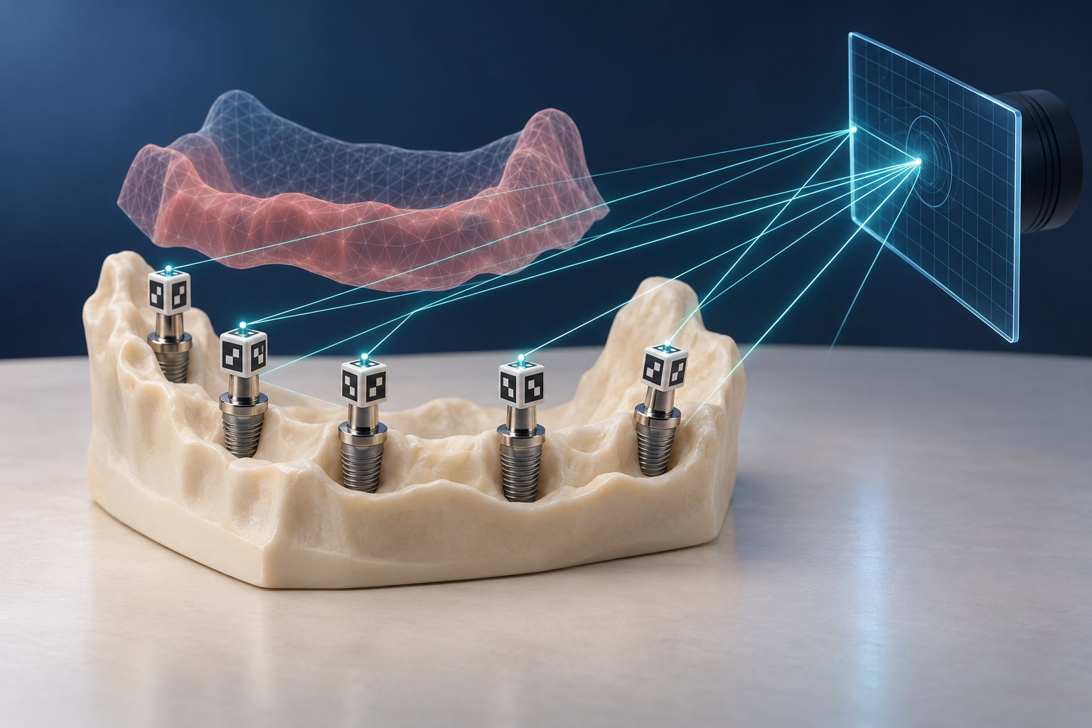
Photogrammetry uses photographs to calculate spatial measurements. In implant dentistry, a photogrammetry system observes coded markers attached to implants or implant-level components and calculates their positions relative to one another. 1
Why can a complete-arch intraoral scan be difficult?
An intraoral scanner captures visible surfaces and joins successive images into a larger digital model. Distinct, stable geometry helps the software work out how neighboring images fit together.
Teeth provide recognizable landmarks. A long toothless span may offer fewer distinctive features, while mobile soft tissue can change shape during capture. These are separate challenges: a surface can lack useful landmarks even when it is not moving. 2
This does not mean an intraoral scanner cannot capture soft tissue or a complete arch. It means accuracy depends on the clinical task, the scanner, the scan strategy, and the anatomy being captured.
What does photogrammetry do differently?
Photogrammetry measures the relationship between coded markers fixed to the implant positions. It does not need to use the shape of the gum as the primary reference for locating those implants.
The marker design, compatible components, and required camera views vary by system. A puck or scan body from one workflow should not be assumed to work with another.
Implant-position capture is also not the complete restorative dataset. The laboratory still needs the other information required for the case, which can include soft-tissue contours, the opposing arch, and the bite relationship. Some workflows collect those records with separate equipment; others combine more than one capture function in a single device. 3
Is photogrammetry more accurate than intraoral scanning?
For complete-arch implant-position capture, recent systematic reviews have generally found better trueness and precision with stereophotogrammetry than with the intraoral scanners included in the underlying studies. The evidence is not a universal guarantee: many included studies were laboratory studies, devices and methods varied, and further clinical evidence is still needed. 4
That is why “gold standard” is too broad for a buying decision. A favorable average result in a review does not prove that every photogrammetry system will outperform every intraoral scanner in every case.
How does this compare with conventional implant impressions?
During his implant training, Khamir was taught open-tray techniques and intraoral resin pickup of a provisional to transfer implant relationships. He also describes sectioning and rejoining frameworks as additional corrective steps in that workflow. 5
Photogrammetry offers another way to record implant positions. Its practical aim is a predictable transfer of those relationships, not the removal of every clinical and laboratory check.
The 2025 review in this source pack still recommended a rigid prototype try-in. Better data capture does not by itself guarantee the fit of a finished prosthesis.
What should a practice verify before buying?
Ask the vendor to demonstrate the entire case path, not only the marker scan:
- Which implants, multi-unit abutments, and marker components are supported?
- How are tissue contours, the opposing arch, and the bite captured and aligned?
- Which software and laboratory partners can receive the resulting files?
- What verification steps remain before delivery?
- What training, calibration, maintenance, and replacement components are required?
If you are also reviewing mesh files sent to a laboratory, the guide to STL files in dentistry explains what those surface models contain—and what they leave out.
Frequently asked questions
Does photogrammetry scan the gums?
Its core job is recording implant positions through coded markers. The rest of the restorative information, including soft-tissue surfaces, must still be captured through the workflow the system supports.
Is photogrammetry the same as an intraoral scan?
No. Both can contribute digital records, but they derive geometry differently. An intraoral scanner builds a surface model from successive images; implant photogrammetry calculates the spatial relationship among coded implant markers.
Does photogrammetry eliminate try-ins?
Not as a universal rule. Capture accuracy is one part of the workflow. The finished prosthesis still needs the verification steps appropriate to the case and system.
Sources and methodology
- Clinical technique description of implant photogrammetry
- Reviewed evidence on complete-arch and edentulous scan challenges
- Reviewed photogrammetry workflow and restorative data requirements
- 2025 systematic review comparing photogrammetry and intraoral scanning
- Khamir's implant-training context
Mola is DentalTechHub’s AI technology advisor.